


Today, let us explore the application of artificial intelligence in the diagnosis and treatment of pulmonary nodules:
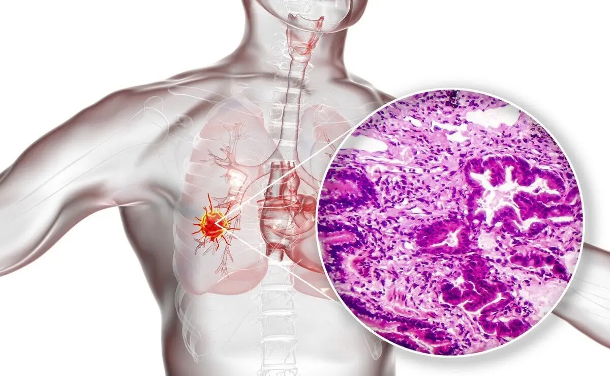
Early-stage lung cancer often lacks typical clinical symptoms and presents only as pulmonary nodules on CT imaging.Low-dose CT is currently the primary screening modality for early lung cancer.Low-dose CT screening for lung cancer has been shown to reduce lung cancer–specific mortality. Broader adoption of lung cancer screening programs has significantly increased pulmonary nodule detection rates, markedly increasing the workload for imaging departments in assessing these nodules.
Recently, the Expert Consensus on the Application of Artificial Intelligence in the Diagnosis and Treatment of Pulmonary Nodules (2022 Edition) (hereinafter the “Consensus”) was formally published in the Chinese Journal of Lung Cancer, China’s first consensus on AI applications in pulmonary nodule diagnosis and treatment. This article elaborates on specific AI applications in pulmonary nodule care in light of the Consensus.
Current status and limitations of pulmonary nodule diagnosis
Studies show that among pulmonary nodules detected by low-dose spiral CT screening, the malignancy rate is only 10%–20%. In the National Lung Screening Trial (NLST), for nodules ≥4 mm in diameter, the false-positive rate after three rounds of low-dose spiral CT screening exceeded 96.4%.
Current guidelines recommend positron emission tomography–computed tomography (PET-CT), endobronchial ultrasound-guided transbronchial needle aspiration (EBUS-TBNA), or transthoracic needle aspiration (TTNA), among other approaches, which have limited sensitivity.
In pulmonary nodule diagnosis, AI can effectively distinguish small nodules from non-nodules, reduce false-positive rates, and increase nodule detection rates.Incorporating 3D texture features of pulmonary nodules, clinical information, and CT image data into a support vector machine model for lung cancer prediction can improve radiologists’ diagnostic sensitivity and specificity.
Expert consensus:
The role of artificial intelligence in pulmonary nodule identification
However, AI sensitivity for subsolid pulmonary nodules is lower; even at the highest sensitivity setting, at most 50% of subsolid nodules can be detected.
Beyond detection, AI can also calculate pulmonary nodule volume and estimate volume doubling time. AI-based volume measurement has high reproducibility, especially for nodules with maximum diameter <10 mm, offering clear advantages over manual diameter measurement.
Expert consensus:
AI has major advantages in assisting physicians with pulmonary nodule identification and is of important value in judging benign versus malignant nature during follow-up (consensus strength: unanimous).
AI has a relatively high false-negative rate for subsolid nodule detection; manual reading confirmation is still needed to reduce missed diagnoses (consensus strength: near-unanimous).
The role of artificial intelligence in differential diagnosis of benign and malignant pulmonary nodules
According to prior reviews, four deep learning models for benign–malignant classification trained and validated on different database types achieved accuracies of 79.5%–93.6%, while models trained and validated on the same database type achieved 68%–99.6%—already relatively high diagnostic accuracy for pulmonary nodules.
In clinical practice, the main advantages of AI-assisted imaging classification of pulmonary nodules include rapidly providing benign–malignant judgments, reducing radiologists’ workload, improving diagnostic efficiency, and highlighting suspicious imaging feature regions for radiologists—helping reduce misclassification.
Expert consensus:
AI technology can provide adjunctive reference for clinical diagnosis in distinguishing benign from malignant pulmonary nodules, but its accuracy cannot yet replace human judgment (consensus strength: unanimous).
Lung cancer diagnostic technologies that fuse multimodal information can achieve more precise diagnostic performance (consensus strength: near-unanimous).
Some scholars have proposed guiding the extent of surgical resection based on intraoperative frozen section results; however, concordance between intraoperative frozen section and postoperative paraffin pathology for lung adenocarcinoma subtypes is insufficient, with sensitivity for micropapillary and solid components of only 37% and 69%, respectively.New methods are urgently needed to assist diagnosis before surgery and guide subsequent treatment.
Traditional CT interpretation largely judges invasiveness based on nodule features.In real clinical practice, however, physicians of different ranks and experience differ in understanding these imaging features and in discrimination ability. Traditional image feature analysis also involves complex procedures, strong human factors, and insufficient feature specificity—affecting interpretation accuracy.
Studies show that AI can effectively distinguish whether ground-glass–predominant lung adenocarcinoma on imaging is invasive disease, providing a clinical basis for early diagnosis and individualized treatment。
Expert consensus:
Relying on deep learning and memory, AI can accurately extract influential microfeatures of pulmonary nodules. With advantages of being noninvasive, capturing tumor heterogeneity, and reproducibility, it holds promise for grading and predicting invasive subtypes of early lung adenocarcinoma presenting as ground-glass nodules, informing clinical decisions. Multicenter, high-quality datasets and prospective randomized controlled trials are still needed for further validation (consensus strength: unanimous).
The role of artificial intelligence in integrated judgment of serial pulmonary nodule follow-up data
In another study, dynamic AI assessment of two imaging examinations of pulmonary nodules also improved lung cancer diagnostic rates. In addition, AI systems can help guide follow-up strategy formulation for pulmonary nodules.
Expert consensus:
In serial follow-up data, AI can help assess changes in pulmonary nodule volume and morphology, providing reference on doubling time and morphologic change to individualize follow-up intervals; specific applicable scope warrants further study (consensus strength: near-unanimous).
The role of artificial intelligence in pulmonary nodule surgical planning (medical 3D reconstruction)
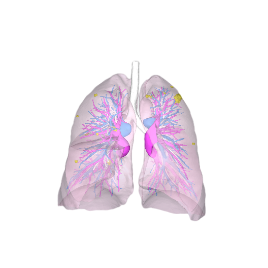
Meanwhile, AI-enabled medical 3D reconstruction can improve surgical success rates and achieve precise resection, especially for preoperative simulation and planning of sublobar resection. Combining visualization with AI methods can help physicians precisely define the extent and surgical pathway for segmentectomy and wedge resection—represented by companies such as Sailner Digital Medical.
Expert consensus:
AI-based medical 3D reconstruction technology is of important significance for improving surgical safety and accuracy (consensus strength: unanimous).