


Jian Zhiheng, Li Jiayan, Ding Xuan, Wu Kaihua, Li Shixue, Chen Haidong, Chen Gang
Department of Neurosurgery, Zhuhai People's Hospital (Zhuhai Hospital Affiliated with Jinan University), 519000
Corresponding author: Chen Gang, Email: jhy_501@163.com
Medicine has entered the era of precision care. Rapid advances in computing have driven progress in medical imaging and computer-assisted surgery, laying the foundation for more precise operations. Among these,medical 3D models and image-based 3D reconstruction play an important role in precise brain tumor surgery. They provide visual maps of spatial relationships and deliver stereoscopic, visualized information to the surgeon, supporting approach design, tumor resection, and protection of brain function. Substantial research has already analyzed the value of this technology in brain tumor surgery[1-4]. Most studies, however, rely on single-modality 3D reconstruction—MRI or CT alone—and therefore cannot display all brain structures relevant to surgery. Multimodal image fusion and 3D reconstruction register and fuse different modalities such as CT and MRI, then reconstruct them in 3D so that the skull, tumor, normal brain tissue, vessels, and nerves appear together in one stereoscopic image. Compared with single-modality reconstruction, this approach better shows spatial adjacency among intracranial structures and is more useful for planning brain tumor surgery and choosing the approach. In recent years, the Department of Neurosurgery at Zhuhai People's Hospital has applied preoperative multimodal image fusion and 3D reconstruction in selected brain tumor patients. Here we report the practical use, outcomes, and lessons learned, in the hope of offering useful experience for clinical colleagues.

I. Materials and Methods
1. General data: Forty-six patients with brain tumors who underwent craniotomy in the Department of Neurosurgery, Zhuhai People's Hospital, from October 2019 to September 2020 were enrolled, including 17 males and 29 females; age (42.2±14.5) years, range 13–77 years. Tumors were supratentorial in 30 cases (cerebral convexity 20, skull base 6, sellar region 4) and infratentorial in 16 (brainstem 1, cerebellum 4, petroclival/cerebellopontine angle/craniocervical junction 11). This study was approved by the Medical Ethics Committee of Zhuhai People's Hospital (approval No.: ZY.No20201001b06011231).
2. Image data acquisition: (1) CT was performed with a GE Revolution CT (128-slice) scanner (USA); slice thickness 2 mm. (2) MRI was performed with a Philips 3.0T Achieva TX MR scanner (Netherlands). Sequences included non-contrast and contrast-enhanced 3D T1 (TE 3.9 ms, TR 8.4 ms, matrix 256×256, FOV 256 mm×256 mm, slice thickness 1 mm), 3D T2 (TE 265 ms, TR 2500 ms, matrix 256×256, FOV 256 mm×256 mm, slice thickness 1 mm), 3D T2 fluid-attenuated inversion recovery (FLAIR) (TE 325 ms, TR 4800 ms, matrix 256×256, FOV 256 mm×256 mm, slice thickness 1 mm), contrast-enhanced MR venography (CE-MRV) (TE 1.62 ms, TR 4.7 ms, matrix 256×256, FOV 256 mm×256 mm, slice thickness 1 mm), and contrast-enhanced MR angiography (CE-MRA) (TE 1.62 ms, TR 4.7 ms, matrix 256×256, FOV 256 mm×256 mm, slice thickness 1 mm).
3. Image import, registration/fusion, and 3D reconstruction: Preoperative CT and MRI DICOM data were imported into GE AW4.4 workstation software (USA) and processed as follows: (1) Different MRI sequences and CT images were registered and fused with the ImageFusion module, using characteristic anatomic landmarks (e.g., globe centers, pituitary fossa, torcular Herophili, mastoid tip, and the apex of the digastric groove) as reference points—3–5 points per patient. (2) The Object Creation module automatically segmented brain tissue, vessels, and nerves and completed automatic 3D reconstruction; tumor location was manually outlined with Smart Brush; skull reconstruction was performed automatically with the 3D-Restruction module. (3) The surgical position was simulated to display and inspect the fused reconstructions of skull, brain, vessels, tumor, and nerves, with transparency adjusted for each structure. Finally, based on the fused 3D digital images, the surgeon designed the approach and surgical plan.
4.Evaluation of clinical value: referring to Oishi et al.[5] grading criteria for the value of image fusion in preoperative planning for microvascular decompression, this study likewise used surgeon consistency grades to assess the value of preoperative multimodal image fusion and 3D reconstruction for brain tumor surgery: after surgery, the primary surgeon and assistant each rated every case. If their ratings differed, the primary surgeon's judgment prevailed. Effects were graded into three levels: (1) Outstanding value: the fused 3D digital images were essential guidance for completing the procedure, and reviewing 2D images alone could not achieve the intended surgical outcome; (2) Valuable: the intended outcome could also be achieved from 2D images, but the fused 3D digital images provided clearer, more intuitive display of fine structures, helping reduce collateral injury and assisting surgery; (3) No value: surgery could be completed as planned from 2D images alone, without multimodal fusion and 3D reconstruction.
5. Assessment of surgical outcomes and complications: All patients underwent postoperative cranial CT and brain MRI using the same standards as preoperative imaging, and multimodal image fusion and 3D reconstruction were repeated from the postoperative studies. Pre- and postoperative 3D digital images were compared to analyze the extent of tumor resection and approach-related complications.

II. Results
1. Multimodal image fusion and 3D reconstruction: All 46 patients completed pre- and postoperative multimodal fusion and 3D reconstruction. Fused multimodal images could be rotated freely, and surface structures rendered transparent or semi-transparent to reveal internal structures of interest. The 3D digital images clearly showed tumor size, location, and shape, as well as anatomic relationships with surrounding structures—for example, 3D reconstructions of superficial and deep veins and arteries clearly demonstrated relationships between tumor and vessels, and complex tumor–normal tissue boundaries and relationships with vessels and skull could be observed in a single image. In all 46 patients, fused 3D digital images were essentially consistent with original preoperative imaging and intraoperative findings, and clearly displayed anatomic information relevant to the surgical approach. Typical fused 3D digital images are shown in Figures 1–4.
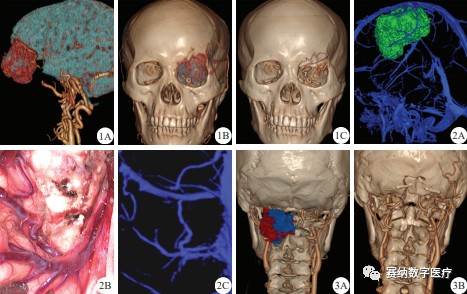
1A: Image fusion and 3D reconstruction of brain tissue/tumor/vessels;
1B, 1C: Pre- and postoperative image fusion and 3D reconstruction of skull/tumor/vessels;
2A: Image fusion and 3D reconstruction of draining veins with a giant frontoparietal meningioma;
2B: Intraoperative view of the Rolandic vein in a giant frontoparietal meningioma;
2C: Image fusion and 3D reconstruction of the Rolandic vein;
3A: Preoperative; 3B: Postoperative
Figure 1. Multimodal image fusion and 3D reconstruction in a complex cranio-orbital communicating tumor
Figure 2. Spatial relationship between draining veins and brain tumor after multimodal image fusion and 3D reconstruction
Figure 3. Pre- and postoperative multimodal image fusion and 3D reconstruction of skull/tumor/vessels in a schwannoma at the craniocervical junction
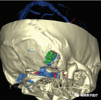
Figure 4
Multimodal image fusion and 3D reconstruction before surgery for a vestibular schwannoma
2. Clinical value ratings: Among 46 patients, the primary surgeon and assistant agreed on the value of preoperative multimodal fusion and 3D reconstruction in 45 cases and disagreed in 1 cerebellar hemangioblastoma (primary surgeon: outstanding value; assistant: valuable); the primary surgeon's rating was adopted. Overall, 36 cases were rated outstanding value, 8 valuable, and 2 no value.
3. Surgical outcomes and complications: Gross-total resection under the microscope was achieved in 43 patients and partial resection in 3 (1 brainstem metastasis from breast cancer, 1 intracranial invasion of external auditory canal squamous cell carcinoma [craniofacial communicating tumor], and 1 optic canal meningioma). No approach-related complications occurred. Postoperative pathology: glioma 9, meningioma 21, metastasis 4, vestibular schwannoma 5, pituitary adenoma 2, trigeminal schwannoma 1, hemangioblastoma 1, cavernous hemangioma 1, craniocervical junction schwannoma 1, and cholesteatoma 1. Thirty-four patients were followed after discharge for 3–6 months; MRI showed no tumor recurrence.

III. Discussion
Understanding the anatomy of a brain tumor and its relationship to surrounding structures before surgery is critical for precise brain tumor surgery. Conventional single-modality CT or MRI no longer meets the needs of precise neurosurgery. An imaging technique that clearly, intuitively, and comprehensively shows the tumor and its relationships to surrounding vessels, nerves, and brain tissue—as well as skull structures related to the approach—is required. Multimodal image fusion registers and overlays images of the same tissue or organ acquired from the same or different modalities using computer technology, producing integrated image information[6]. With this technology, neurosurgeons can fuse large imaging datasets to reconstruct visualized anatomic structures in 3D and clarify tumor–surrounding relationships from multiple viewpoints; and, based on skull, brain, vessel, and nerve information related to the approach, they can more precisely design the incision and bone flap and more effectively avoid eloquent cortex and critical vessels[7-8].In addition, visualized 3D reconstruction of anatomic structures can deepen neurosurgeons' understanding of complex intracranial anatomy and tumor pathology, providing greater assistance during resection[9].
We performed preoperative multimodal image fusion and 3D reconstruction for 46 brain tumor patients and found that the fused 3D digital images clearly displayed tumor size, location, shape, and relationships with surrounding structures, effectively guiding approach selection and precise resection. Key lessons include:
(1) In complex skull-base tumor surgery, this technology clearly shows the relationship between tumor and skull. For example, in one patient with an orbito-cranial communicating optic canal meningioma, it clearly demonstrated the extent of tumor invasion into the anterior skull base, periorbital bone, and optic canal.
(2) When formulating the preoperative plan, fused 3D digital images can accurately localize relationships between vessels and tumor. Regarding draining veins, among 20 convexity tumor patients we found draining veins immediately anterior to the tumor within the bone window in 5 cases, immediately posterior in 6, crossing the tumor surface in 3, and only tumor without closely related draining veins in 6; accordingly, we could deliberately avoid and protect draining veins during craniotomy. In one giant convexity meningioma at the frontoparietal junction, multimodal fusion and 3D reconstruction clearly showed the tumor anteroinferior to the Rolandic vein and delineated the entire vein and its relationships to the superior sagittal sinus, small tributaries, and tumor—information that traditional 2D images could not clearly provide—thereby helping reduce the risk of venous injury during resection. Regarding arteries, we also observed arteries displaced by tumor in 7 cases and encased by tumor in 3; clarifying these relationships helped the surgeon anticipate the position of encased arteries intraoperatively, enabling more confident piecemeal resection and more careful arterial protection.
(3) For tumors at the craniocervical junction and related arterial protection, multimodal fusion and 3D reconstruction also greatly aid preoperative planning. In one craniocervical junction schwannoma, the technology clearly showed relationships among the tumor, the right vertebral artery, and bony structures including cervical spinous processes, intervertebral foramina, and the foramen magnum region, revealing destruction of the C2 foramen and complete encasement of the transverse, atlantal, and foramen magnum segments of the vertebral artery—information that allowed more deliberate tumor resection while preserving the artery.
(4) For deep brain tumors, approach-related injury must be considered, and bone-flap design must both avoid venous injury and facilitate resection. In one vestibular schwannoma patient, careful review of the fused 3D digital images allowed precise localization of the surface projections of the transverse and sigmoid sinuses when designing the bone flap, avoiding injury to these sinuses during milling.
(5) In many low-grade gliomas, the boundary between superficial tumor and normal brain is unclear, making intraoperative identification and localization difficult and often requiring intraoperative electrophysiologic monitoring to avoid eloquent-area injury. Mert et al.[10] previously used multimodal fusion and 3D reconstruction to assist low-grade glioma resection; the fused tumor images accurately showed relationships with adjacent brain and helped reduce the monitored area and operative time. Unfortunately, our own experience with multimodal fusion and 3D reconstruction for low-grade gliomas with little MRI enhancement remains limited, and further practice and summary are needed.
The two patients rated "no value" both had frontal convexity meningiomas <3 cm in diameter, with clear MRI T2 borders, moderate homogeneous enhancement, no critical vessels inside or around the tumor, location in a relatively silent area, and low surgical difficulty; thus multimodal fusion and 3D reconstruction added little. In the one case of rating disagreement—a right cerebellar hemangioblastoma—the feeding artery was already relatively clear on preoperative 2D images, so the assistant rated it valuable; the primary surgeon felt 2D images did not clearly show the relationship between the nodule and feeding artery, whereas the fused 3D digital images displayed this spatial relationship very clearly and were important during fenestration and tumor exploration, and therefore rated outstanding value.
In summary, preoperative multimodal image fusion and 3D reconstruction provide abundant visualized information and have broad prospects for guiding incision design, approach selection, precise tumor resection, and protection of brain function. Limitations remain: for vessel reconstruction, although we used multiple sources such as 3D phase-contrast MRV, CE-MRA, or time-of-flight MRA, no single method reconstructs tiny vessels satisfactorily; for tumor reconstruction, 3D T1WI is commonly used and works well when enhancement is marked, but reconstruction is difficult when enhancement is mild or absent. We believe these shortcomings will continue to improve as related technologies advance.
Conflicts of interest
All authors declare that they have no conflicts of interest

References:
[1] Nandish S, Prabhu G, Rajagopal KV. Multiresolution image registration for
multimodal brain images and fusion for better neurosurgical planning[J]. Biomed J, 2017, 40(6): 329-338. DOI:10.1016/j.bj.2017.09.002.
[2] Hoffmann N, Weidner F, Urban P, et al. Framework for 2D-3D image fusion of
infrared thermography with preoperative MRI[J].Biomed Tech (Berl), 2017, 62(6): 599-607. DOI: 10.1515/bmt-2016-0075.
[3] Neuberger I, Hankinson TC, Meier M, et al. Utility of imagefusion software in
identifying shunt malfunction[J]. Childs NervSyst, 2020, 36(4): 749-754. DOI: 10.1007/s00381-019-04385-9.
[4] Inoue HK, Nakajima A, Sato H, et al. Image fusion forradiosurgery, neurosurgery and hypofractionated radiotherapy[J].Cureus, 2015, 7(3): e252. DOI: 10.7759/cureus.252.
[5] Oishi M, Fukuda M, Hiraishi T, et al. Interactive virtual simulationusing a 3D
computer graphics model for microvasculardecompression surgery[J]. J Neurosurg, 2012, 117(3): 555-565.DOI: 10.3171/2012.5.JNS112334.
[6] Hirano T, Ichikawa K, Wanibuchi M, et al. Accuracy of computedtomography-magnetic resonance imaging image fusion using a phantom for skull base surgery[J]. J Neurosurg Sci, 2019. DOI:10.23736/S0390-5616.19.04621-6.
[7] Li J, Zhang Y, Le H, et al. Application of multimodal image fusiontechnology in brain tumor surgical procedure[J]. Translational Neuroscience and Clinics, 2016, 2(4): 215-226. DOI: 10.18679/CN11-6030_R.2016.035.
[8] Yin Tengkun, Wang Shousen. Research progress on venous circulation assessment related to parasagittal meningioma and its surgical value[J]. Chinese Journal of Neuromedicine, 2018, 17(7): 738-741. DOI: 10.3760/cma.j.issn.1671-8925.2018.07.019.
Yin TK, Wang SS. Venous assessment and surgical strategy inparasagittal meningiomas[J].Chin J Neuromed, 2018, 17(7): 738-741. DOI: 10.3760/cma.j.issn.1671-8925.2018.07.019.
[9] Sato M, Tateishi K, Murata H, et al. Three-dimensionalmultimodality fusion imaging as an educational and planning toolfor deep-seated meningiomas[J]. Br J Neurosurg, 2018, 32(5):509-515. DOI: 10.1080/02688697.2018.1485877.
[10] Mert A, Buehler K, Sutherland GR, et al. Brain tumor surgerywith 3-dimensional surface navigation[J]. Neurosurgery, 2012, 71(2 Suppl Operative): ons286-294; discussion ons294-295. DOI:10.1227/NEU.0b013e31826a8a75.
Excerpted from Chinese Journal of Neuromedicine, November 2021, Vol. 20, No. 11
Original editor: Liu Kai
Some images in this article are sourced from the Internet