


Expert Interview:
Prof. Zhuang Jian, Cardiovascular AI and 3D Technology Laboratory, Guangdong Provincial People’s Hospital
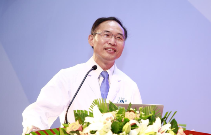
Brief Biography
Zhuang Jian, M.D., Chief Physician, Professor, and doctoral supervisor, is a national selectee of the New Century National Hundred, Thousand and Ten Thousand Talent Project and a young and middle-aged expert with outstanding national contributions. He currently serves as Director of the WHO Collaborating Center for Research and Training in Cardiovascular Diseases, Director of the Guangdong Cardiovascular Center, Chief Expert in Cardiac Surgery at Guangdong Provincial People’s Hospital, Director of the Guangdong Provincial Key Laboratory of Structural Heart Disease in South China, and Director of the Cardiovascular AI and 3D Technology Laboratory at Guangdong Provincial People’s Hospital. He previously served as Dean of the School of Medicine at South China University of Technology, President of Guangdong Provincial People’s Hospital, Immediate Past Chair of the Chinese Society for Thoracic and Cardiovascular Surgery of the Chinese Medical Association, Chair of the Cardiovascular Surgery Branch of the Guangdong Medical Doctor Association, Council Member of the Asian Society for Cardiovascular and Thoracic Surgery, Editorial Board Member of The Annals of Thoracic Surgery (USA), and Associate Editor-in-Chief of the Chinese Journal of Thoracic and Cardiovascular Surgery. He has long been engaged in surgical treatment and systematic prevention research for severe and complex congenital heart disease (CHD), pioneering multiple new procedures in China for complex CHD surgery, and pioneering the integrated application of 3D printing, virtual reality, and mixed reality in CHD surgical adjunctive care. He led the establishment of China’s first provincial comprehensive CHD prevention and treatment network and guided clinical and research teams to elevate complex CHD surgical correction at Guangdong Provincial People’s Hospital / Guangdong Cardiovascular Institute to internationally advanced levels.
In 2018 he received one Guangdong Provincial Science and Technology Progress First Prize, and in 2019 one 11th Soong Ching Ling Pediatric Medicine Award. Over the past decade he has led 10 national and provincial/ministerial key science and technology projects, including those under the 11th and 12th Five-Year Plans, published more than 300 papers—including over 60 SCI papers as corresponding or first author in leading journals such as European Heart Journal and Circulation—obtained more than 10 patents, and led the formulation of China’s first standards for fetal echocardiography and prenatal counseling for congenital heart disease.
Complex congenital heart disease (CHD) is highly heterogeneous and extremely difficult to diagnose and treat. Complex CHD comprises many disease types; each major category subdivides into multiple subtypes, and even within the same subtype the shape and course of a given structure—and the spatial relationships among structures—can differ completely. This makes anatomy, pathophysiology, and hemodynamic changes far more complex than in simple CHD.
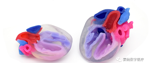
In surgical clinical practice, common adjunctive examinations include echocardiography, computed tomography (CT), magnetic resonance imaging (MRI), angiography, chest radiography, electrocardiography, and cardiac catheterization. These modalities provide direct 2D imaging, partially reconstructed 3D imaging, and directly or indirectly measured hemodynamic parameters. Operators must rely on imagination and prior experience to reconstruct anatomy mentally and then interpret pathophysiology and hemodynamics. When applied to highly individualized complex CHD, these approaches severely tax spatial imagination, biomechanical understanding, and memory, while also introducing subjectivity that increases uncertainty in diagnosis, treatment strategy, and outcomes—hindering quantitative assessment of the relationship between strategy and effect.
Pediatric cardiac surgery at Guangdong Provincial People’s Hospital began in the 1980s, when congenital heart disease (CHD) treatment in China was also just getting started. To overcome surgical challenges, Prof. Zhuang Jian made many pioneering attempts—he was among the first in China to apply multiple new procedures to complex CHD surgery, including originating the “arterial switch with ventricular septal defect fenestration repair,” giving new life to many children who previously could only wait and perish.
Through years of effort by pediatric CHD surgeons represented by Dr. Zhuang Jian, China has gradually aligned with—and in some areas reached—international leadership in pediatric CHD surgery.
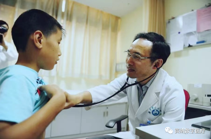
Looking at global CHD surgery development, although skilled use of conventional examinations and procedures has greatly improved outcomes for complex CHD, major challenges remain for many complex diseases and operations. First, surgical difficulty is high: many procedures can be performed well only by a few top experts at home and abroad, with limited disseminability, long learning curves for young surgeons, and high operative cost. Second, outcomes still have substantial room for improvement: even when complex operations succeed, they often involve long operative times, multiple or large incisions, and many postoperative complications. How to deliver individualized care for the strong heterogeneity of complex CHD, further improve outcomes, reduce difficulty, and increase disseminability has become the pursuit of CHD surgery today.
Hospital–Industry Collaboration: Seeking Breakthroughs
Drawing on years of clinical experience, Prof. Zhuang realized that new imaging-assisted diagnostic and treatment modalities are key to these goals. In 2014, his team partnered with Xidian University to apply for a National Natural Science Foundation Key Project.
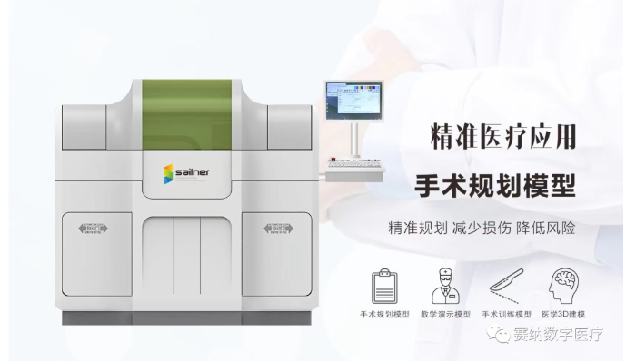
“Research on Key Enabling Technologies for Complex CHD Surgery Assistance Based on Multimodal Image Fusion and Visualization” was funded, initiating clinical research on virtual reality (VR). The project ultimately produced multiple outcomes, including a first-generation VR display system for preoperative diagnosis and planning; a cardiac multimodal image fusion technique with a related patent, “Cardiac CT–TEE Registration Method Based on Valve Alignment and Probabilistic Maps”; and new methods for hemodynamic prediction in selected diseases, with a related patent, “Device and Method for Determining Pressure Gradient at Aortic Coarctation from CT Images.” In early 2017, after further investigation, Prof. Zhuang turned to 3D printing technology and promptly contacted China’s leading domestic full-color multi-material digital medical 3D modeling and printing company (Zhuhai Sailner Technology) for technical exchange and collaboration, actively exploring 3D printing applications in congenital heart disease.
After repeated investigation and discussion, Prof. Zhuang decided to begin with several of the most complex CHD types, including pulmonary atresia with ventricular septal defect and major aortopulmonary collateral arteries (PA/VSD/MAPCAs), transposition of the great arteries, and double-outlet right ventricle. After obtaining printed models for selected cases, he intuitively felt that, compared with conventional imaging, these models offer the natural advantage of direct visualization and hold great potential in surgical planning, physician training, and clinician–patient communication—potentially helping address the strong heterogeneity of complex CHD. Taking PA/VSD/MAPCAs as an example, 3D printed models can intuitively show the spatial relationships of great vessels, collateral vessels, and the trachea, enabling the primary surgeon, before sternotomy, to use medical 3D modeling and printing technology to print a cardiac model and clearly explore cardiovascular structures—benefiting surgical planning, intraoperative navigation, and physician training.
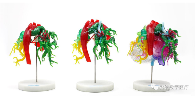
Prof. Zhuang also found that fully and effectively realizing clinical 3D printing faces many challenges. First, domestic and international clinical application standards and guidelines are lacking—which diseases to use it for and how remain to be explored. Second, imaging 3D reconstruction takes too long and relies heavily on experienced imaging physicians, limiting clinical efficiency. Third, material fidelity remains far from adequate for surgical simulation and physician training. Other barriers include the lack of existing fee schedules in the healthcare system and a shortage of interdisciplinary talent who understand both medicine and engineering.
Building a Platform and Implementing in Clinic
To better enable clinical translation and gradually address these barriers, under Prof. Zhuang’s leadership, Guangdong Provincial People’s Hospital and Zhuhai Sailner Technology jointly established the “Cardiovascular Medicine 3D Printing Joint Laboratory” in October 2017—China’s first 3D printing laboratory formed through hospital–research–enterprise collaboration, designed as a lab for communication, integration, and exchange between engineering technology and clinical needs.
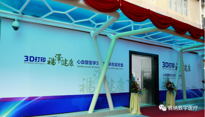
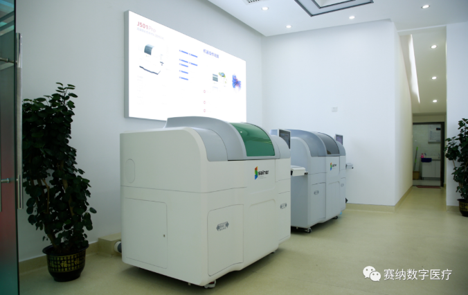
Located inside Guangdong Provincial People’s Hospital, the laboratory greatly facilitates exchange. It houses two of Zhuhai Sailner’s latest domestically developed color multi-material 3D printers. Resident staff include multiple engineers providing professional technical services, as well as physicians and medical students conducting hospital–industry research and clinical application docking.
Shortly after the laboratory’s founding, Prof. Zhuang also actively introduced mixed reality (MR) technology. Together with the continuously upgraded structural heart disease VR-assisted diagnosis and treatment system, the laboratory gained R&D and service capabilities in 3D printing, VR, and MR. After rapid and careful technical integration, in March 2018 the system was formally rolled out for comprehensive clinical use as an adjunctive diagnosis and treatment platform. At the end of 2018, Prof. Zhuang’s team successfully co-applied for and received support under the Guangdong Provincial Key R&D Program, focusing on improving printing technology and materials and vigorously advancing clinical 3D printing.
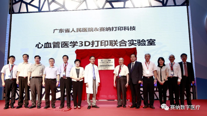
October 2017: Cardiovascular Medicine 3D Printing Joint Laboratory established
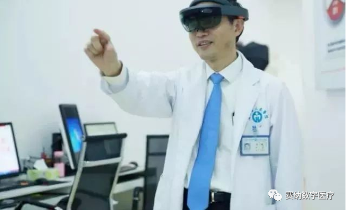
Preoperative surgical planning using 3D printing and virtual reality
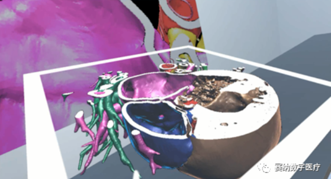
Preoperative surgical demonstration using 3D printing and virtual reality
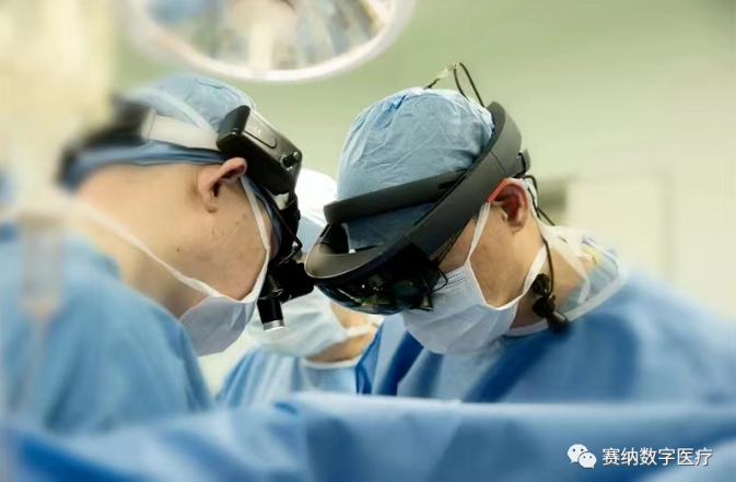
Intraoperative navigation using mixed reality
After clinical implementation, 3D technologies brought many benefits to diagnosis and treatment, and clinical application standards gradually took shape. First, within one week of the laboratory’s setup, all three 3D technologies were applied in the diagnosis and treatment of 10 cases of complex CHD and cardiac tumors—including coronary artery fistula and pediatric left ventricular tumors—with favorable outcomes. The most typical disease was PA/VSD/MAPCAs; for unifocalization surgery of this condition, 3D assistance reduced the incision from the previous “median plus lateral thoracotomy” to a single median incision and effectively shortened operative time. Prof. Zhuang’s team subsequently applied 3D technologies across many complex CHD cases and expanded beyond CHD to hypertrophic cardiomyopathy, great vessel disease, valvular disease, and other structural heart diseases.
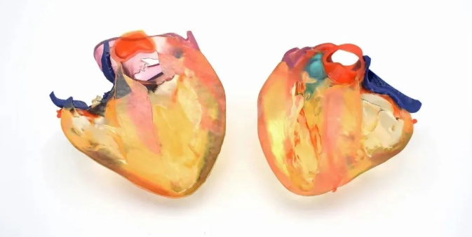
Sailner FLX920 series ultra-soft material 3D printing for a hypertrophic cardiomyopathy case
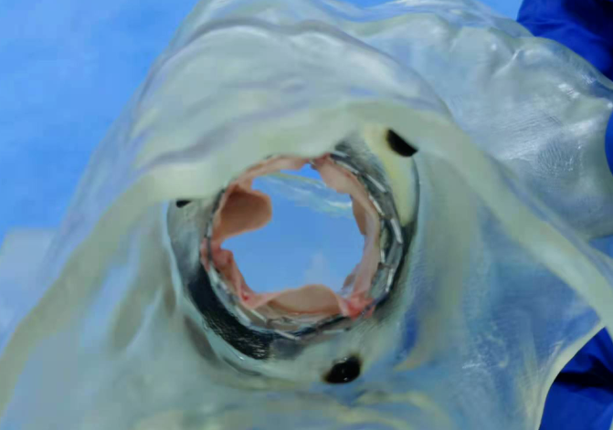
Sailner FLX910T series ultra-flexible material 3D printing of valvular disease for surgical simulation
By the end of 2021, clinical applications of 3D technologies in cardiac surgery had exceeded 2,000 cases. Second, after accumulating case experience and rigorous cost accounting, three medical fee standards for cardiac 3D technology applications applicable in Guangdong Province were established—clearing compliance barriers and laying a realistic foundation for sustainable use. Third, through continuous experience, basic standards for 3D reconstruction and application across multiple diseases gradually formed. Overall standards include color coding by anatomical site, applicable disease scope, and selection among 3D printing / VR / MR; disease-specific standards cover image data requirements, reconstruction site selection, print site selection and segmentation, and material selection by site. Taking PA/VSD/MAPCAs as an example, beyond reconstruction requirements, a selective printing protocol was developed—printing only pulmonary arteries/collaterals, aorta, and trachea/bronchi—meeting clinical practicality and timeliness while reducing printing cost. Fourth, long-term, multi-case clinical use cultivated many interdisciplinary professionals—including surgeons more fluent in 3D technologies and engineers more attuned to clinical needs—advancing both medicine and engineering.
Expanding Research and Promoting Application
To improve the timeliness of 3D reconstruction, since 2018 Prof. Zhuang has deeply collaborated with AI medical imaging experts and, in 2019, full-time recruited young postdoctoral AI talent, forming a larger hospital–industry research team. The laboratory was upgraded to the “Cardiovascular AI and 3D Technology Laboratory” with an expanded research scope. A key focus—intelligent modeling and diagnosis of CHD CT images—has advanced AI denoising and image recognition, reducing noise while shrinking image volume to facilitate subsequent segmentation. To date, intelligent modeling and diagnosis of 17 common CHD types can largely be achieved, with mean image segmentation DICE of 73% and diagnostic accuracy of about 86%, comparable to junior cardiovascular imaging physicians. AI-based 3D technologies are also improving and innovating traditional care models. For example, in 2018 Prof. Zhuang’s team conducted remote guidance of a thoracoscopic atrial septal defect repair using AI + 5G: preoperative denoising and reconstruction produced a precise 3D CT model; VR was used to record a detailed surgical planning video specifying port positions; a physical model was printed for real-time guidance; and the surgery was successfully completed.
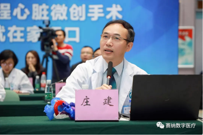
Prof. Zhuang Jian providing 5G remote surgical guidance
The team is now deploying related AI algorithms into modeling software to replace part of manual modeling and improve efficiency, while researching how to further raise AI modeling and diagnostic accuracy for large-scale clinical application. Using AI, the team has also built a prediction model for postoperative pulmonary venous obstruction after total anomalous pulmonary venous connection repair based on raw CT images and clinical parameters, with prediction performance clearly superior to ordinary models—a novel attempt at structural heart disease data mining.
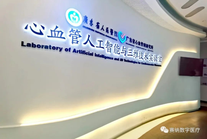
The Cardiovascular AI and 3D Technology Laboratory today
Accessibility of AI and 3D technologies is essential for clinical use and for sustainable hospital–industry–enterprise research. Since the laboratory’s founding, accessibility of AI and 3D technologies across all specialties at Guangdong Provincial People’s Hospital has greatly increased. Beyond cardiac surgery, physicians in pulmonology, orthopedics, hepatobiliary surgery, plastic surgery, and other disciplines have obtained technical support through the laboratory and actively pursued innovative clinical applications.
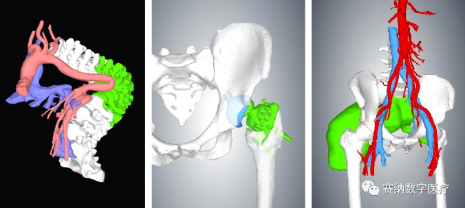
Clinical applications in spine, joint, and bone tumor fields

Clinical applications in ENT, breast, and hepatopancreatobiliary fields
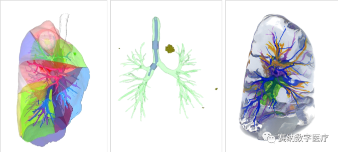
Clinical applications in pulmonary surgery
For example, pulmonology has developed more intuitive pulmonary nodule localization methods based on 3D technologies—including watershed analysis localization based on 3D reconstruction, 3D-printed bone guides, and mixed-reality fluoroscopic localization—helping refine wedge resection to the subsegmental level while reducing preoperative radiation. These efforts have also partly addressed application standards, interdisciplinary talent development, and fee schedule formulation, advancing new technologies into clinical practice for more patients. Effective application and timely clinical feedback have likewise driven continuous technology advancement; pulmonary vessel intelligent reconstruction accuracy is reportedly already very high. This fully demonstrates that new technologies and clinical application can mutually reinforce and develop healthily.
Medical 3D modeling and printing, moving from engineering into the clinic, effectively helps physicians “rebuild” patients’ cardiac health. This translation is the result of close collaboration between medical and technical professionals. Looking ahead, we hope more talent from both engineering and medicine will join hospital–industry research, bringing more new technologies into real clinical practice to solve clinical problems, make innovative—even revolutionary—changes toward medical homogenization and better outcomes, and contribute more to health in China and worldwide.
Acknowledgments: The original interview text is from the Additive Manufacturing Medical Devices Committee