



Pulmonary atresia with ventricular septal defect (PA/VSD) is a rare and highly complex congenital heart disease, accounting for approximately 2.5%–3.4% of all congenital heart defects. The main malformations include a large ventricular septal defect, overriding aorta, and atresia of the pulmonary artery or right ventricular outflow tract. According to the internationally used Castaneda classification, the condition is divided into four types; Type IV denotes the absence of true pulmonary arteries, with pulmonary blood supply derived entirely from major aortopulmonary collateral arteries (MAPCAs), and is the most severe form.
The most common symptoms in affected children are cyanosis and hypoxemia. Some neonates appear well at birth, then develop life-threatening hypoxemia as the ductus arteriosus closes. If the infant survives without fatal ductal closure in the neonatal period or heart failure from large MAPCAs, a circulatory balance may allow survival into adolescence. During this period, chronic progressive cyanosis leads to polycythemia and, over the long term, may cause cerebral embolism and brain abscess, along with marked clubbing of the fingers and toes.
Surgical treatment of Type IV PA/VSD typically requires unilateral or bilateral lateral thoracotomy first to identify and control the major MAPCAs, followed by conversion to median sternotomy for unifocalization of bilateral MAPCAs into new left and right pulmonary arteries, reconstruction of the right ventricular outflow tract connecting the right ventricle to the new pulmonary arteries, and repair of the ventricular septal defect either in the same stage or in a staged procedure. The operation is extremely demanding and high-risk; only a very small number of hospitals in China currently offer it.
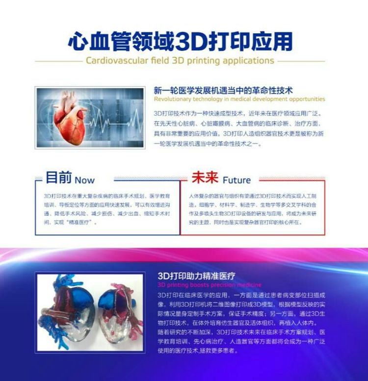
President Zhuang Jian of Guangdong Provincial People’s Hospital, who also serves as Chair of the Chinese Society for Thoracic and Cardiovascular Surgery, leads a congenital heart surgery team with the largest domestic experience in treating Type IV PA/VSD. President Zhuang and his team are also studying the clinical application of advanced 3D printing technology, especially for surgical treatment of complex congenital heart disease, such as3D-printed heart models。
On October 11, 2017, Sailner Digital Medical and the Cardiovascular Medicine 3D Printing Joint Laboratory of the Provincial People’s Hospital Cardiovascular Research Institute were formally established—China’s first laboratory of its kind, with major significance for the diagnosis and treatment of cardiovascular disease.
Case 1: a 4-year-old with Type IV PA/VSD had cyanosis of the lips, hands, and feet since birth. His parents sought care at many major hospitals, all of which deemed surgery impossible. Fortunately, they found Chief Physician Wen Shusheng of the Department of Cardiac Surgery at Guangdong Provincial People’s Hospital. After reviewing the work-up, Dr. Wen told the family that young patient Z could undergo surgery, and that medical 3D printing of a complex congenital heart model could improve surgical success and reduce risk—news that greatly relieved the parents.
After admission, preoperative preparations proceeded in an orderly fashion. Director Huang Meiping of Radiology completed modeling based on cardiac CT data, and the 3D printing laboratory then performed3D printing of the heart model. Before surgery, President Zhuang, Dr. Wen, and colleagues carefully studied the heart model, clarifying the course of each collateral vessel and its relationship to surrounding tissues. Surgery began with a right lateral thoracotomy; guided by the spatial relationships shown on the 3D model, Dr. Wen quickly located several major collateral vessels—completing in only 1 hour a process that usually takes several hours. The team then converted to median sternotomy, where President Zhuang completed MAPCA unifocalization and right ventricular outflow tract reconstruction. The operation went smoothly; the child’s cyanosis resolved, and recovery was good at discharge. Several months later, young patient Z will return for ventricular septal defect repair, after which he can expect near-normal cardiac function, life expectancy, and quality of life.
Case 2: a 6-year-old with the same Type IV PA/VSD had also consulted multiple major hospitals before presenting to the Provincial People’s Hospital. Building on the prior case, President Zhuang and Dr. Wen decided that, given the high fidelity and precise localization of the 3D-printed model, they could attempt to find the collateral vessels through a median incision alone—saving the time of lateral thoracotomy opening/closing and re-draping, and reducing injury to the lung and respiratory muscles to aid recovery. This proved correct: during surgery, Dr. Wen accurately located all four MAPCAs in only 45 minutes. After assessing that the child’s condition was suitable, President Zhuang performed MAPCA unifocalization, right ventricular outflow tract reconstruction, and ventricular septal defect repair for one-stage radical correction, shortening the overall operative time by 5 hours compared with previous practice. The girl recovered well, was weaned from ventilatory support on postoperative day 3, and was expected to be discharged soon.