


What is medical 3D reconstruction?
Human medical 3D reconstruction technology is characterized chiefly by medicine and information engineering, reconstructing three-dimensional anatomical structures of the human body in a virtual reality environment. The medical images needed for modern human medical research and clinical diagnosis and treatment are diverse—such as X-ray, CT images, nuclear medicine images, magnetic resonance (MRI) images, ultrasound images, and various electronic endoscopic images—but these images can only provide two-dimensional views of the body interior. Faced with 2D images, clinicians can only estimate lesion size and shape from experience and mentally conceive the three-dimensional geometric relationship between the lesion and surrounding tissues, which creates great inconvenience for diagnosis and treatment.
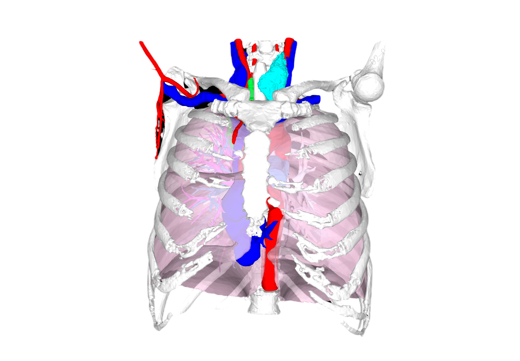
Three-dimensional visualization of medical images presents human body information obtained from digital imaging equipment such as CT and MRI as intuitive 3D effects on a computer, thereby providing structural information unobtainable by traditional means. Its task is to reconstruct three-dimensional images with computers and display lifelike stereoscopic views of human organs on screen. Through human–computer interaction, medical professionals can conveniently rotate, translate, section, and window the reconstructed images, refine preoperative assessment, and perform visual simulated surgery. This enables medical professionals to more fully understand the nature of the lesion and the three-dimensional structural relationships of surrounding tissues, helping them make accurate diagnoses and formulate correct surgical plans, thereby improving the accuracy and scientific basis of diagnosis and treatment.
Surgical case
For example, Ms. A noticed swelling on the right side of her neck more than a decade ago. She was examined at a hospital and diagnosed with nodular goiter, and underwent right thyroidectomy as advised. However, five years ago she found the other side of her neck swelling as well. Examination showed not only left thyroid enlargement, but also that residual thyroid tissue on the right from the previous resection had extended into the thoracic cavity, with an overall diameter exceeding ten centimeters in a dumbbell shape, compressing adjacent thoracic organs. Without surgery the consequences would be serious and could even affect breathing. After evaluation, many hospitals concluded that only open thoracotomy could resolve the problem, but the surgery would be highly difficult, highly traumatic, and slow to recover from. For this reason Ms. A later sought care at Guangdong Provincial People’s Hospital.
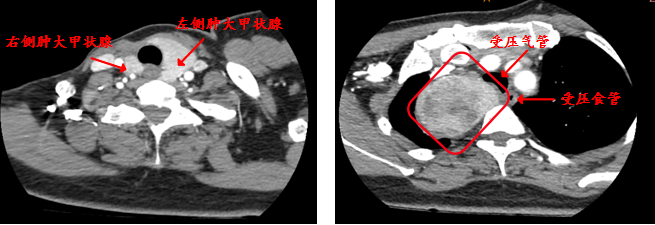
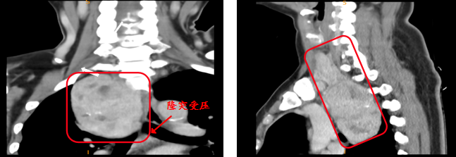
The Sailner Digital Medical team worked with Guangdong Provincial People’s Hospital to perform 3D image reconstruction of the lesion and found that the mass not only encircled the neck and compressed the trachea, but also compressed the right pulmonary vein, tracheal carina, esophagus, and other structures within the chest. Surgery was highly difficult and prone to major bleeding during dissection; postoperatively, tracheal or esophageal perforation or fistula could also occur.
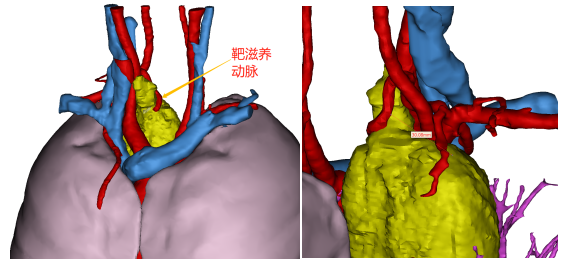
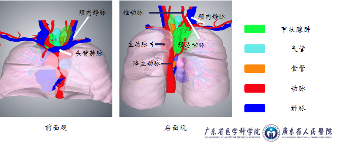
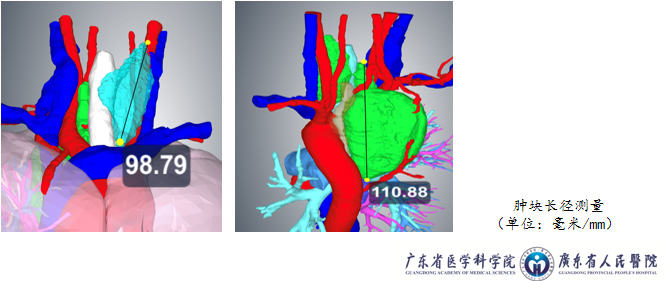
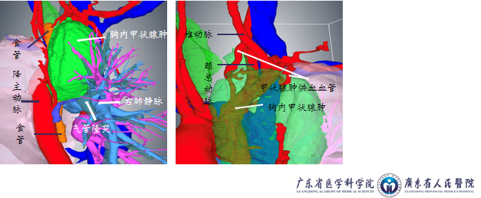
After team discussion and study, the Guangdong Provincial People’s Hospital team found that the cervical and thoracic portions of the right-sided mass were continuous—smaller above and larger below, resembling an eggplant. Through collaborative exploration they ultimately decided that Director Hu Shixiong’s team would first resect the left cervical lesion and free the cervical portion of the right lesion, after which the thoracic surgery teams of Directors Yang Xuening and Zhong Wenzhao would remove the mass via minimally invasive thoracoscopic surgery. Ultimately the surgery was completed smoothly as planned. Ms. A recovered well postoperatively and was discharged after five days—demonstrating the power of teamwork and a direct embodiment of three-dimensional technology applied in medicine.
Medical science popularization
Origins of intrathoracic thyroid tumors—
(1) Thyroid tissue remaining in the mediastinum from the embryonic period later develops into an intrathoracic thyroid tumor;
(2) Originally a cervical thyroid tumor that later descends into the retrosternal space, most commonly in the anterior superior mediastinum, and also seen in the middle and posterior mediastinum.
Clinical features—
The disease is common in people around 50 years of age, predominantly women
Aberrant thyroid often has no clinical manifestations, though some may present with hyperthyroidism
Retrosternal thyroid often presents with cervical goiter, tracheal compression symptoms, or associated hyperthyroidism
Classification of intrathoracic goiter—
Based on imaging findings and clinical symptoms, three types are recognized:
Type I: More than half of the cervical goiter lies retrosternally, with the lower pole reaching the upper border of the aortic arch;
Type II: Nearly the entire enlarged thyroid lies behind the sternum, with the lower pole reaching behind the aortic arch or entering the posterior mediastinum;
Type III: A massive goiter protruding into the thoracic cavity, or accompanied by superior vena cava compression syndrome
Retrosternal thyroid masses are generally benign lesions, but 5%–15% are thyroid malignancies
Key diagnostic points—
Size and shape: Intrathoracic goiter most often presents as a unilateral irregular mass; bilateral cases appear as symmetric or asymmetric saddle-shaped dumbbell masses
On contiguous slices, the mass continues upward with the cervical thyroid and may move up and down with swallowing
Imaging findings—
X-ray: Widened superior mediastinum continuous with the neck; increased density; tracheal compression, deformity, and displacement
CT: Often located anterior or lateral to the trachea, with heterogeneous density, cystic change, hemorrhage, and calcification; calcification is a main feature of intrathoracic goiter. Marked and prolonged enhancement due to iodine uptake
MRI: Long T1 and long T2 signal, heterogeneous, with cystic and necrotic areas; after contrast, the solid portion shows continuous marked enhancement, while cystic and necrotic areas show no enhancement.