


On June 16–17, 2023, the 2023 National Regional Medical Center Medical Science and Technology Forum, hosted by Guangdong Provincial People’s Hospital and Guangdong Provincial People’s Hospital Ganzhou Hospital (Ganzhou Municipal Hospital), opened in Ganzhou with strong momentum. The Guangdong Lung Cancer Research Organization of Guangdong Provincial People’s Hospital convened a symposium on “Fluorescence Laparoscopy–Guided Precise Resection Using the Pulmonary Watershed Analysis Method,” inviting more than 60 thoracic surgery experts from 37 centers for discussion and exploration. Sailner Digital Medical, a company dedicated to medical 3D digital application solutions, also took part in the symposium.
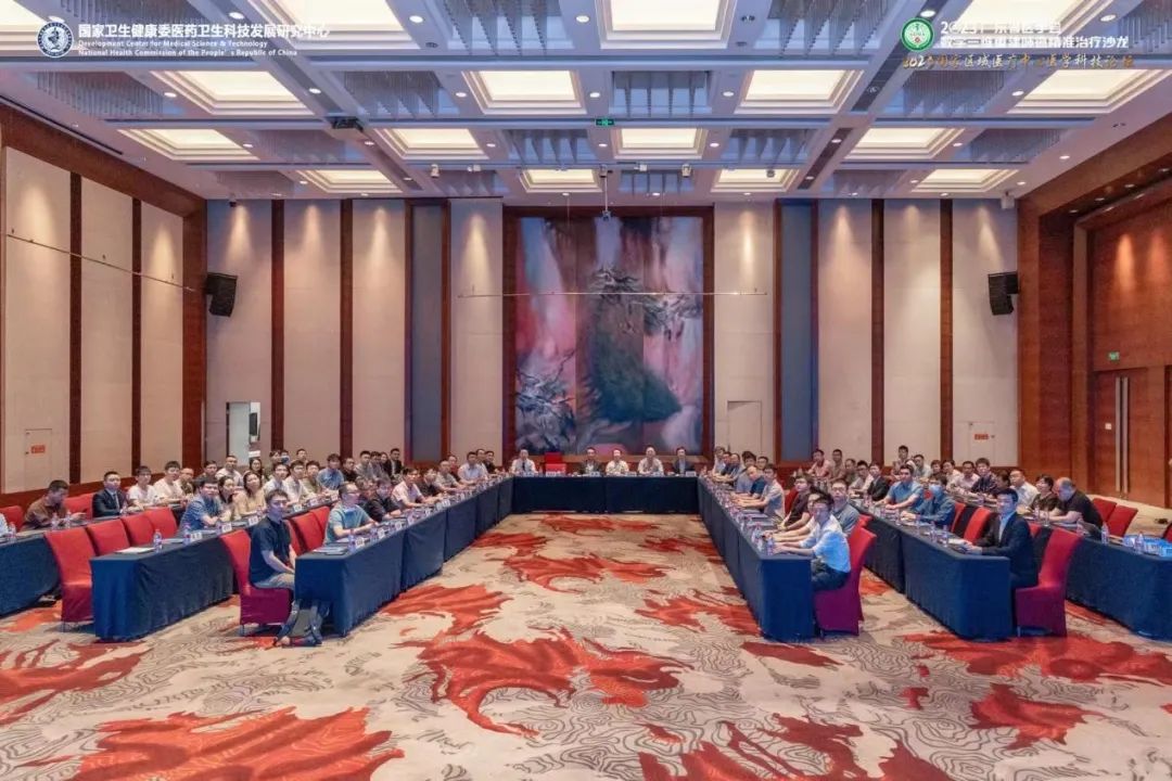 Conference site
 Sailner Digital Medical exhibition booth
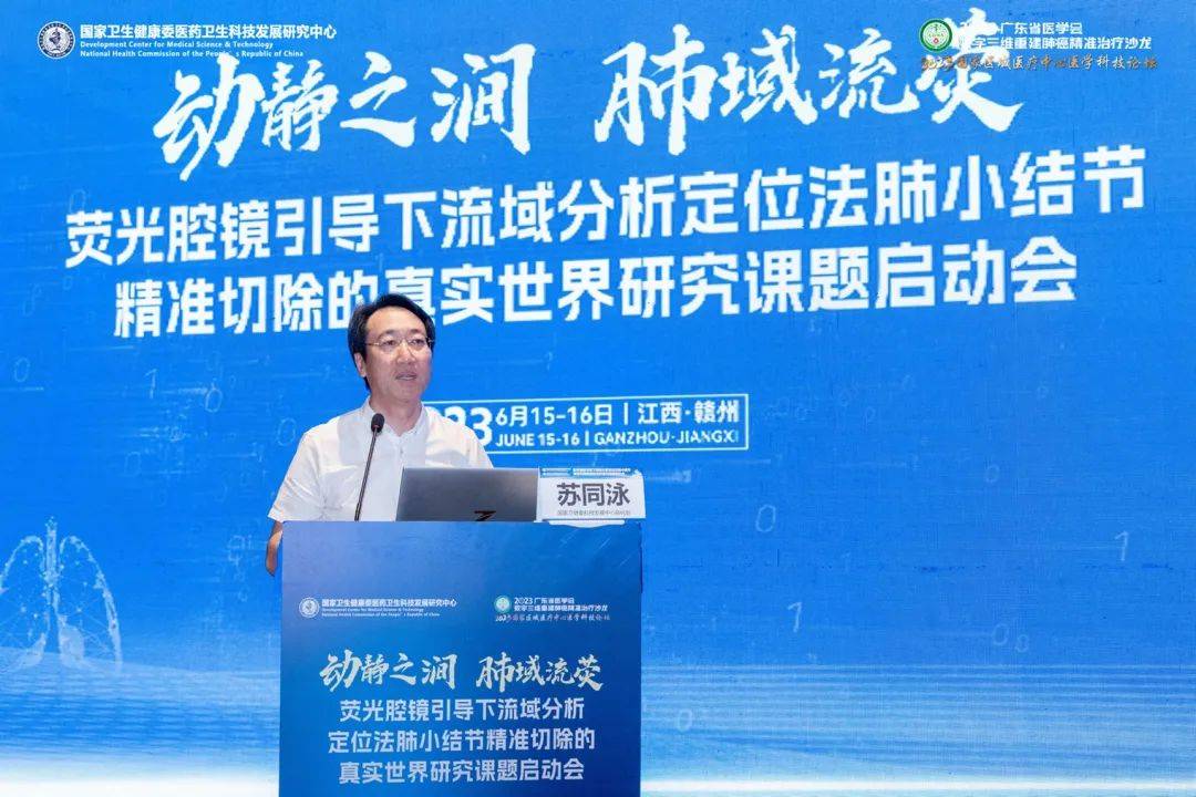 Director Su Tongyong of the National Health Commission’s Research Center for Medical and Health Science and Technology Development delivered remarks and outlined project management requirements
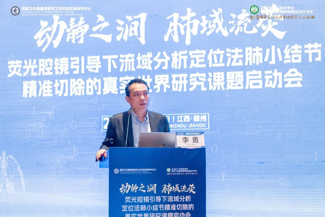 Professor Li Yong, Vice President of Guangdong Provincial People’s Hospital, delivered remarks: the importance of new technology development and clinical translation of research outcomes
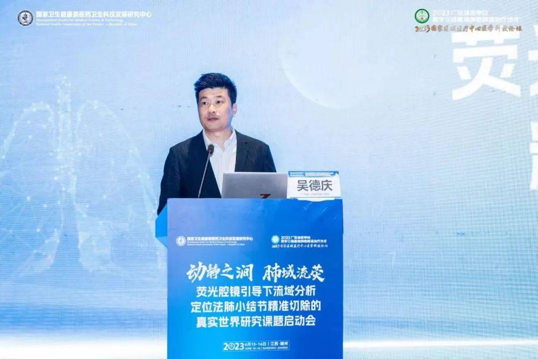 Professor Wu Deqing, President of Guangdong Provincial People’s Hospital Ganzhou Hospital, moderated
I.
Based on CT 3D reconstruction of individualized pulmonary anatomical variations and innovative AI algorithms for true watershed anatomical boundaries, the team led by Director Zhong Wenzhao of the Guangdong Lung Cancer Institute developed a precise pulmonary nodule resection approach—“pulmonary watershed topographic regional resection” with real-time intraoperative navigation. It accurately simulates the watershed of the target artery and vein where the nodule resides, measures the adjacency between the nodule and the watershed, and guides real-time intraoperative localization and resection strategies, achieving innovation in both localization and resection extent. Among these,clinical 3D reconstruction technology was provided by Sailner Digital Medical.
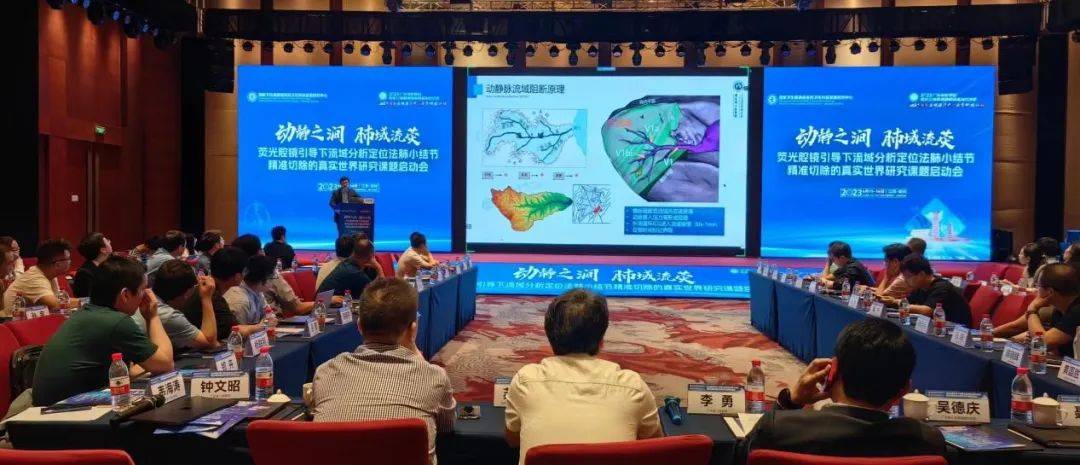 Event site
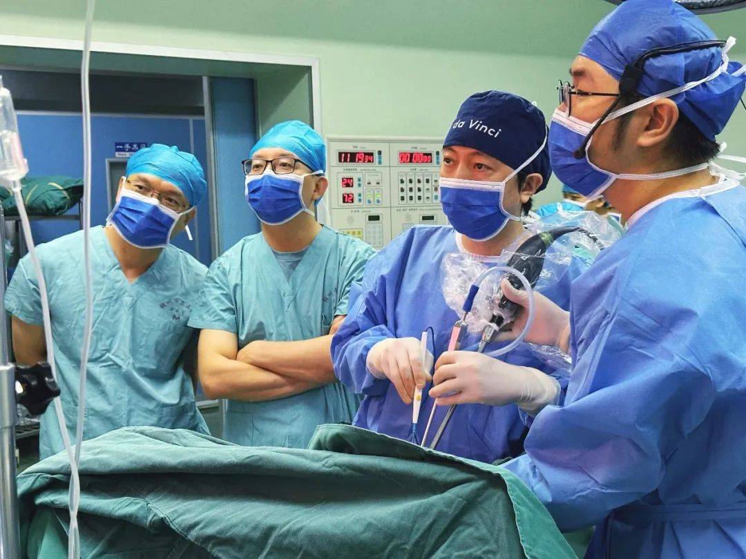 Professor Zhong Wenzhao and Dr. Chen Zihao performing surgery under real-time intraoperative watershed topographic map navigation at Ganzhou Municipal Hospital
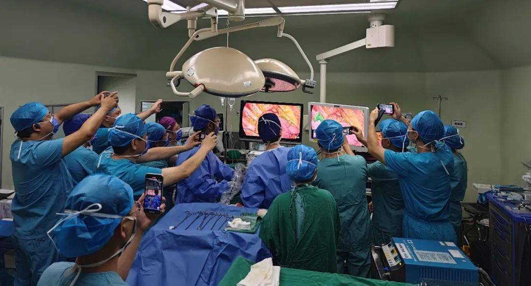 Photo and video recording
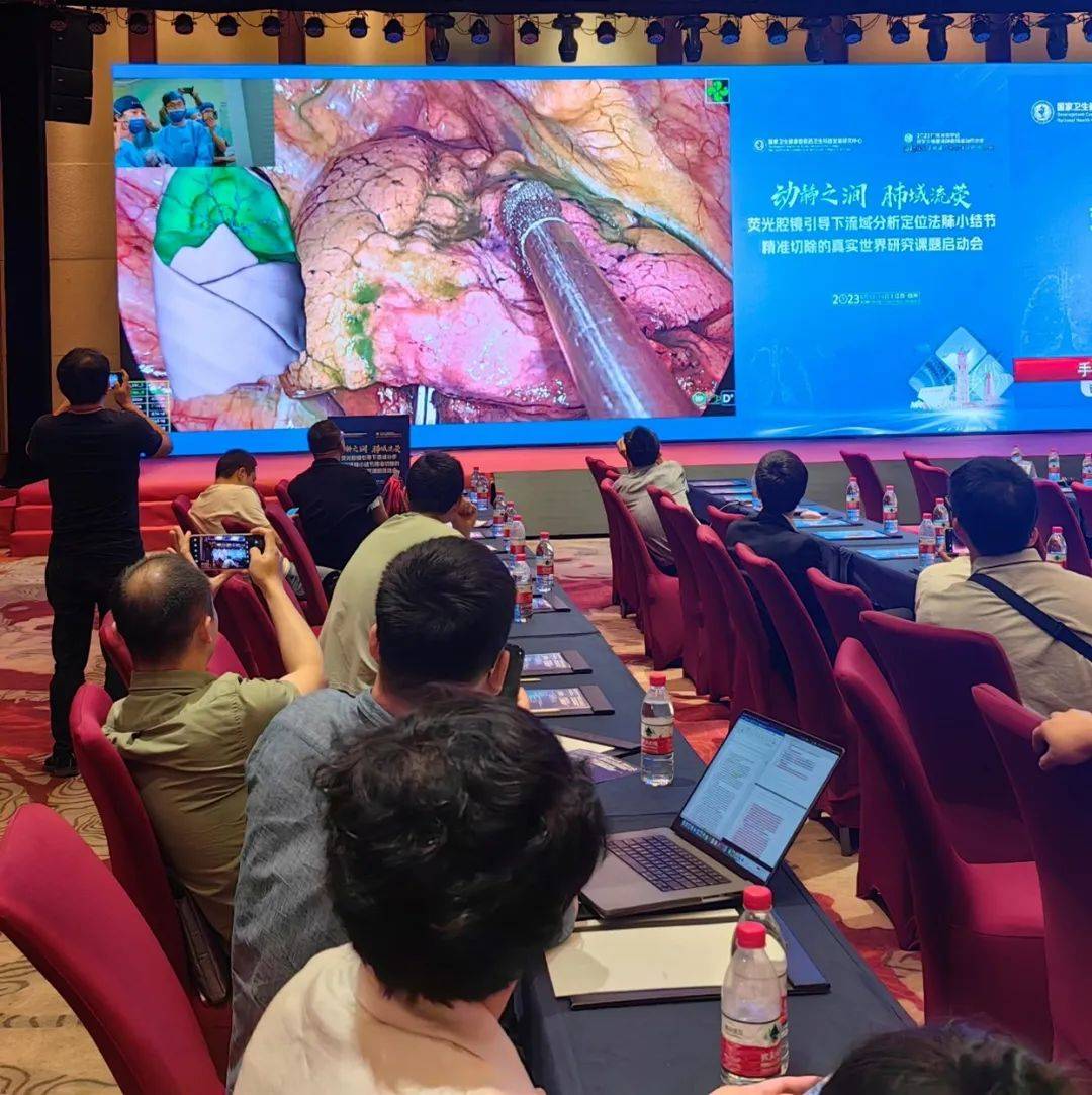 Live broadcast from the Ganzhou venue
II.
Watershed Topographic Map 2.0 uses AI algorithms to simulate the true boundaries of pulmonary vascular watersheds based on individualized pulmonary anatomical variation patterns. Principle of the watershed topographic map: digitally simulate true boundaries using the arterial–venous watershed divide; occluding a branch pulmonary artery dries the watershed blood supply; occluding a branch pulmonary vein causes venous hypertension and blood stasis in the watershed, so peripheral circulation of ICG enters the watershed slowly—thus, after peripheral intravenous ICG injection, a clear watershed boundary appears within 30 seconds to 1 minute. This guides preoperative planning and real-time intraoperative navigation, shortening operative time and reducing risks such as bleeding.
Breakthroughs in clinical practice bottlenecks are mainly reflected in the following four aspects:
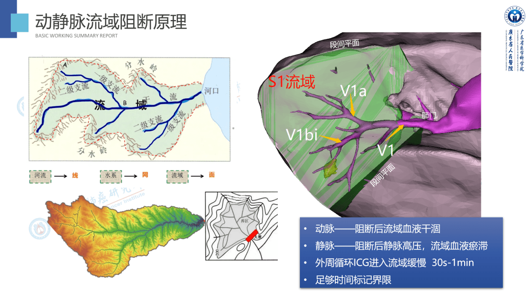 Principle explanation
Breakthroughs in clinical practice bottlenecks are mainly reflected in the following
four aspects:
01 Sand-table–style real-time intraoperative planning
It supports deeper understanding of anatomical variation patterns at segmental boundaries, enables personalized pulmonary watershed topographic maps, and provides sand-table–style strategic guidance for preoperative and intraoperative planning, helping avoid anatomical blind spots in advance. While ensuring margins, it shortens operative time and reduces risks such as bleeding.
02
Enhanced recovery with maximal lung function preservation
Maximally preserves patients’ lung function and supports rapid postoperative recovery. Shortens hospital stays and improves the efficiency of scarce medical resources.
03
Breaking the “restricted zones” of CT localization
It makes minimally invasive resection possible for pulmonary nodules located in conventional CT-guided puncture localization “restricted zones.”
04
Easy to disseminate; accelerating homogenization at primary care
With a short learning curve and ease of dissemination to primary care, it is an effective advanced technique for grassroots settings, accelerating tiered diagnosis and treatment and homogenization of regional care.
III
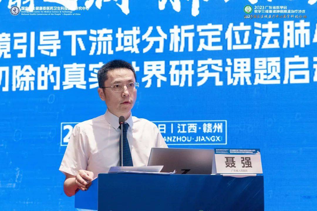 Professor Nie Qiang of the Guangdong Lung Cancer Institute, Guangdong Provincial People’s Hospital, introduced operative details of the watershed method
“Watershed topographic map navigation is a new technique for real-time precise intraoperative localization of pulmonary nodules. The technology has now iterated to version 2.0, which we call the pulmonary watershed topographic map. It enables sand-table–style strategic mapping to design individualized procedures and minimize the extent of lung resection. Based on individualized pulmonary anatomical variation patterns, it also guides preoperative planning and real-time intraoperative navigation, shortening operative time and reducing complications and lung function loss,” said Professor Nie Qiang of the Guangdong Lung Cancer Institute, Guangdong Provincial People’s Hospital. The technique breaks through the “restricted zones” of conventional CT-guided puncture localization and provides a convenient minimally invasive resection option for nodules in special locations.
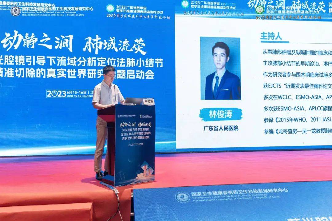 Dr. Lin Juntao of the Guangdong Lung Cancer Institute moderated the session.
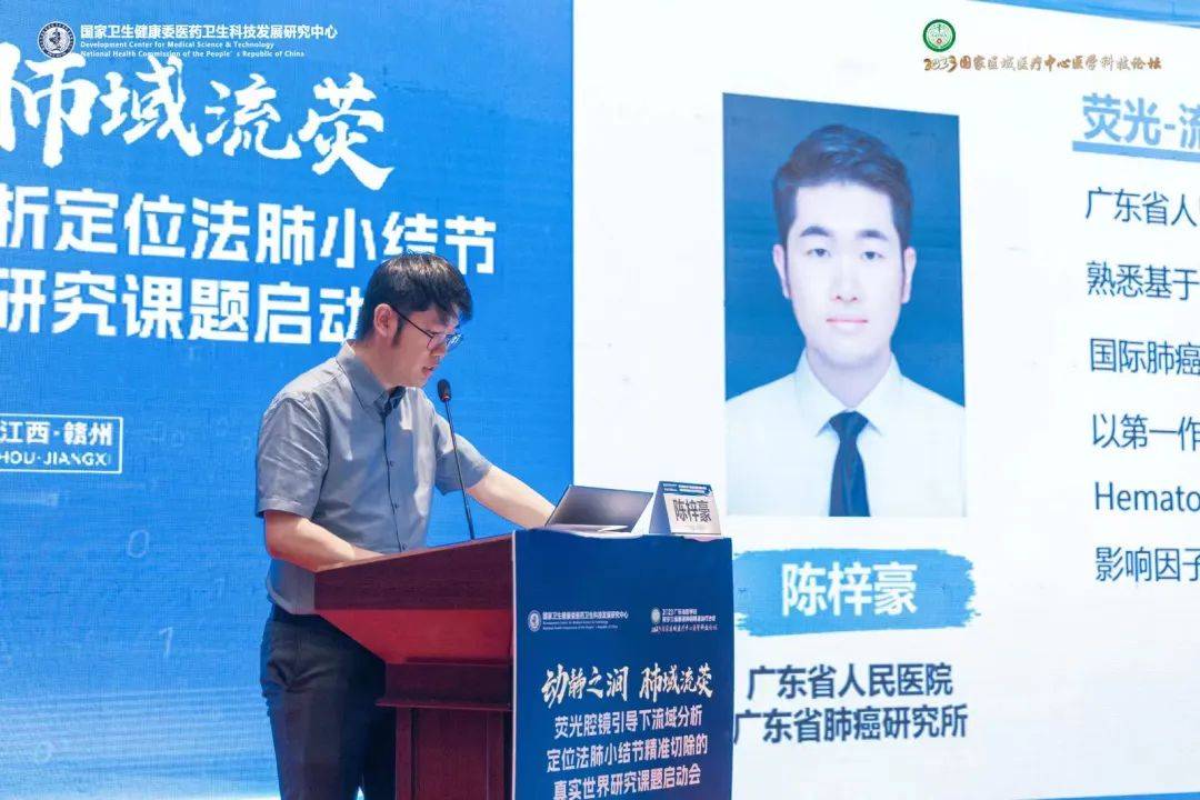 Dr. Chen Zihao of the Guangdong Lung Cancer Institute introduced real-time drawing methods for the watershed topographic map algorithm and the mixed-reality navigation system for lung cancer surgery.
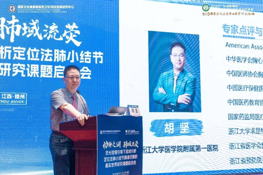 Director Hu Jian, The First Affiliated Hospital, Zhejiang University School of Medicine
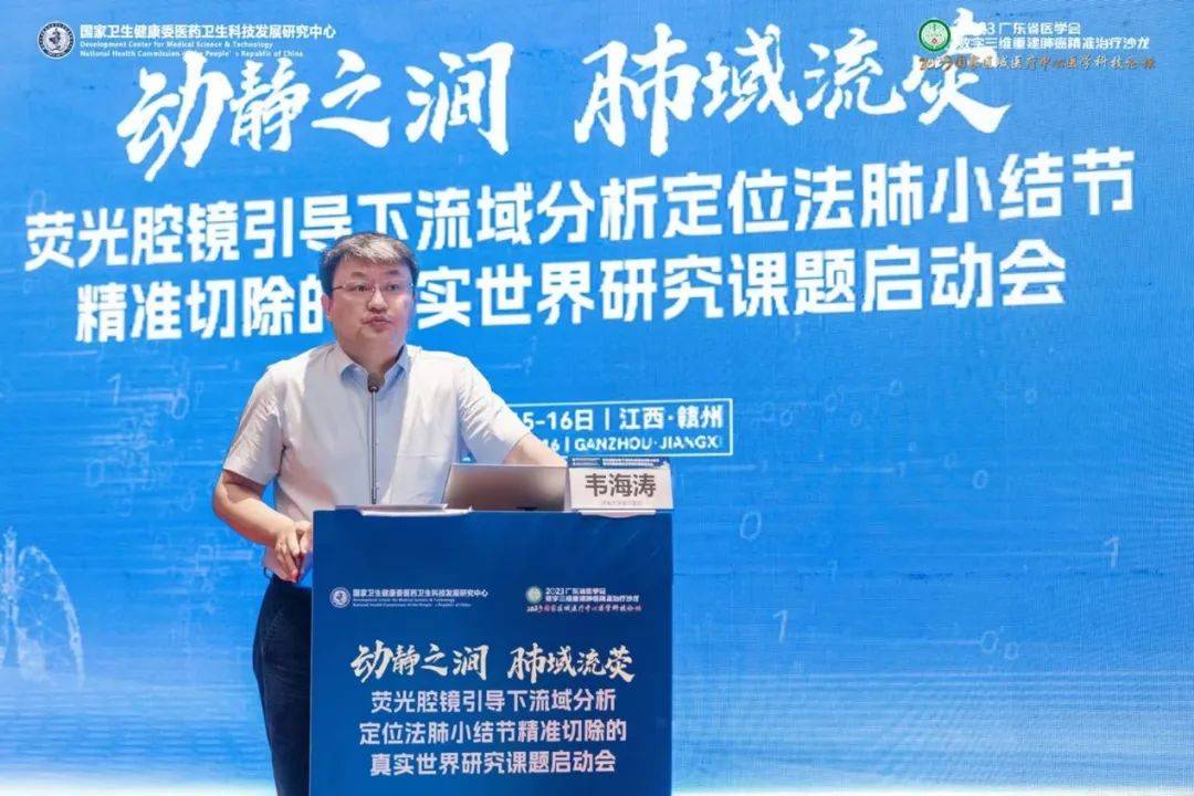 Wei Haitao, Huaihe Hospital of Henan University
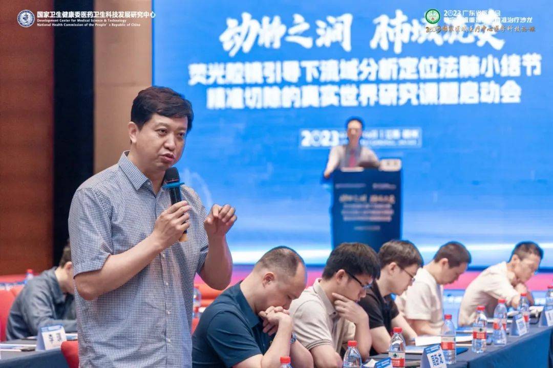 Panel discussion
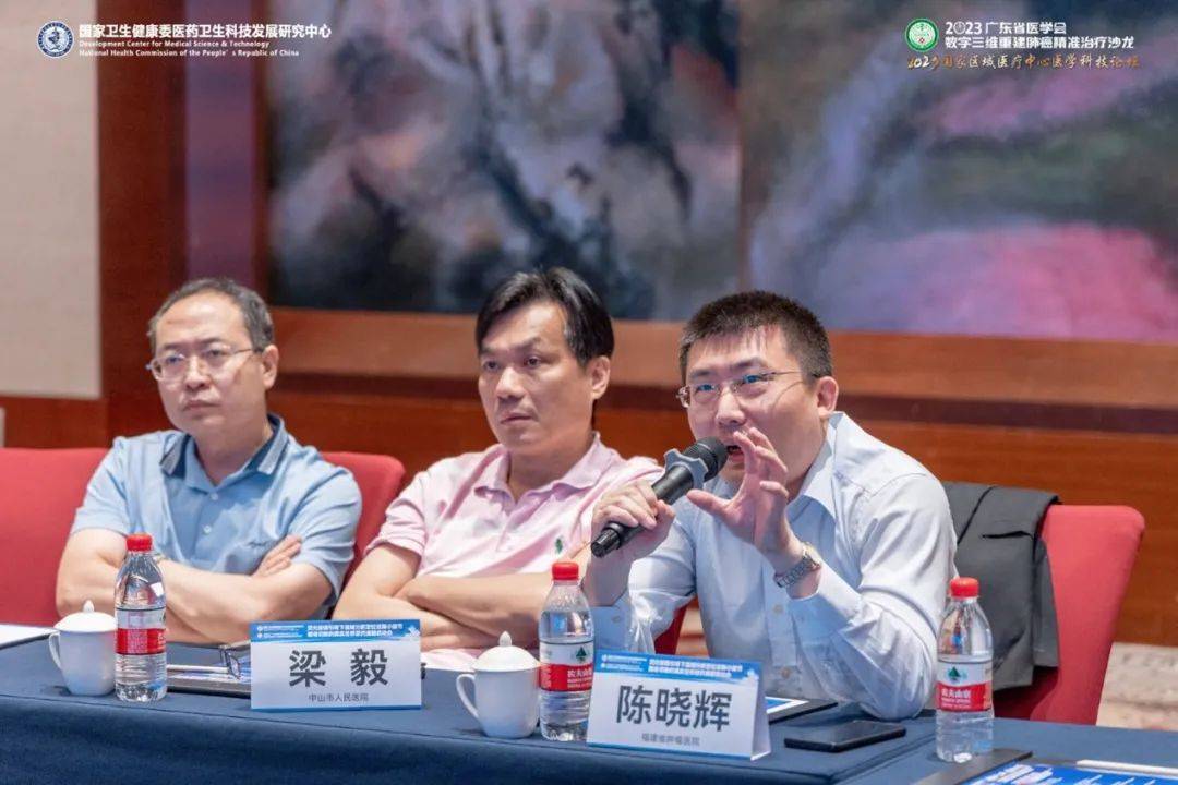 Director Sun Nan of Liaoning Cancer Hospital, Director Chen Xiaohui of Fujian Cancer Hospital, and Director Liang Yi of Zhongshan People’s Hospital joined the discussion.
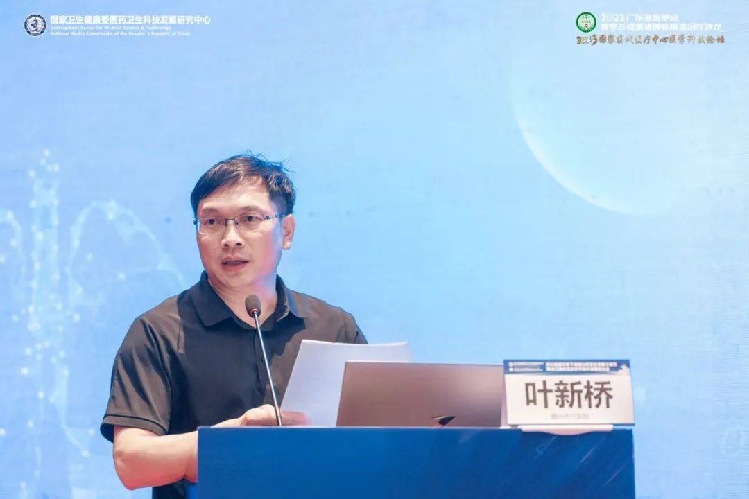 Director Ye Xinqiao of Ganzhou Municipal Hospital moderated the discussion.
IV.
Fluorescence laparoscopy is an important foundation for this technique. Compared with the traditional inflation–deflation method, fluorescence imaging can rapidly, accurately, and clearly display watershed boundaries during surgery, reducing difficulty and shortening operative time, and providing reliable technical support for thoracoscopic anatomical segmentectomy. Opmandy’s fluorescence laparoscope, combined with ICG dosing and fluorescence gain techniques to adjust high-definition visualization time in real time, is a safe and effective approach that merits clinical promotion.
A series of technical innovations and breakthroughs in preoperative planning for sublobar resection under 3D reconstruction guidance and intraoperative watershed analysis localization have reduced patients’ preoperative localization burden and improved the safety and precision of regional resection. In a retrospective analysis of 131 patients, intraoperative blood loss, postoperative drainage rate, postoperative pneumothorax rate, length of stay (days), and pulmonary function loss at 3 months (3–5%) showed no difference versus wedge resection; there were no perioperative deaths; the one-time successful wedge resection rate reached 98%; mean operative time (from incision protector placement to chest closure) was 16.7 minutes. The preoperative planning workflow and technique have a short learning curve and high disseminability, building an information exchange bridge between primary hospitals and provincial hospitals, fostering efficient benign interaction, and contributing to the rollout of the tiered healthcare system.
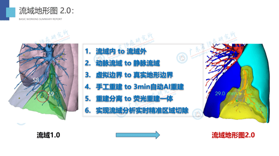 Watershed Topographic Map 2.0