


Approach selection for extended myectomy is influenced by the phenotype of hypertrophic obstructive cardiomyopathy.
The apical approach is an innovative way to improve visualization in this procedure and also a technical challenge. Combined with high-precision technologies such as 3D printing, delivering standardized, precise surgery for patients with hypertrophic cardiomyopathy is a lasting direction for future development.
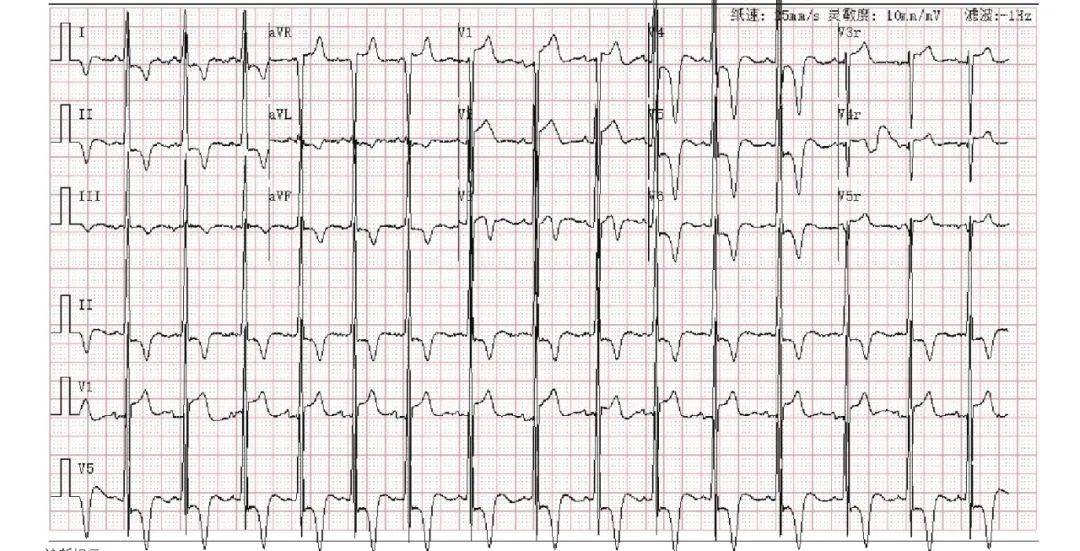
Clinical data A 26-year-old male patient presented to our hospital in December 2021 with “recurrent exertional dyspnea for 10 years, worsening for 1 month.” He reported onset of exertional dyspnea about 10 years earlier without clear precipitating factors, requiring rest after climbing one flight of stairs before relief; he had no chest tightness or chest pain, no dizziness or syncope, and no dyspnea at rest, and received no specific intervention. One month before admission, symptoms worsened, prompting presentation. ECG showed “sinus rhythm, left ventricular hypertrophy, marked ST-segment changes and deep T-wave inversion” (Figure 1). Color Doppler echocardiography suggested “hypertrophic cardiomyopathy, mid-to-lower left ventricular cavity obstruction with a small apical aneurysm; mild mitral regurgitation; reduced left ventricular diastolic function.” He was admitted to our department with a diagnosis of “hypertrophic cardiomyopathy” for further workup and elective surgery.
 Electrocardiogram
Figure 1 Preoperative ECG
ECG shows increased QRS voltage and deep T-wave inversion
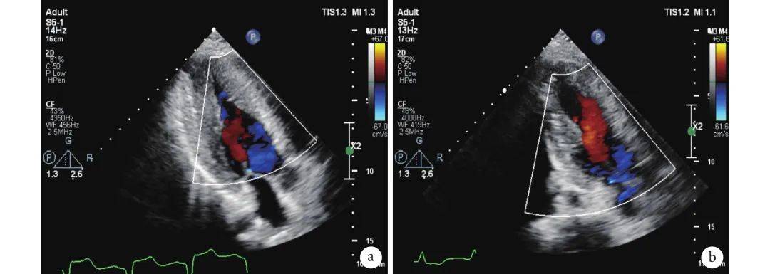
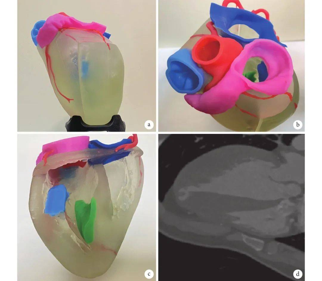
Admission examination: temperature 36.2°C, pulse 66 beats/min, respiratory rate 18 breaths/min, blood pressure 136/72 mm Hg (1 mm Hg = 0.133 kPa); clear breath sounds bilaterally without dry/moist rales or wheezing; heart borders not enlarged, heart rate 66 beats/min, regular rhythm, grade 1/6 diastolic blowing murmur at the left sternal border in the 3rd–4th intercostal spaces; soft abdomen without tenderness or rebound tenderness, liver and spleen not palpable, no extremity edema. Preoperative assessment: diagnosis of “apical hypertrophic cardiomyopathy, ventricular aneurysm, mild mitral regurgitation.” No family history, no history of syncope or amaurosis, and no coronary artery disease risk factors; New York Heart Association (NYHA) class III, EuroSCORE II 0.67%, Society of Thoracic Surgeons (STS) score 0.625%. Transthoracic echocardiography on December 8, 2021: chambers not enlarged, LVEF 70%; septal thickness 13 mm; marked uniform thickening of the mid-to-lower left ventricular myocardium, maximum about 29 mm, with markedly narrowed mid-to-lower cavity adjacent to the papillary muscles (Figure 2a), nearly obliterated; local paradoxical motion at the apex suggesting a small aneurysm; high-velocity flow in the mid-to-lower LV cavity in systole, Vmax = 4.2 m/s, with early-diastolic reverse flow; mild mitral regurgitation, regurgitant area 1.6 cm2; LV volume 39 mL. Preoperatively, 3D modeling and printing were used: cardiac CT 3D images were imported into Mimics for annotation and segmentation, then printed (Figures 3a–b), with structures color-coded (Figure 3c). In the model, the aneurysm (Figure 3d) measured about 18 mm × 5 mm, located on the apical surface and communicating with the ventricular cavity, so the apical incision could be placed over the aneurysm. Myocardial thickness was measured at the base, mid septum, and apex, confirming hypertrophy mainly at the apex (maximum 22 mm) without marked mid-septal stenosis, and no hypertrophied papillary muscles or abnormal muscle bundles connecting to the valve; thus the main resection range was the anterior and posterior septum from the papillary muscle roots to the apex—i.e., the procedure could be completed via an apical approach alone without an aortic approach.
 Echocardiography
Figure 2 Pre- and postoperative transthoracic echocardiography
a: Preoperative echocardiography showing apical hypertrophy and cavity narrowing; b: Postoperative echocardiography showing enlarged cavity
 Sailner Digital Medical 3D-printed model
Figure 3 Patientcardiac 3D-printedmodel and CT images
a: 3D model coronal view; b: 3D model axial view; c: 3D model cross-section, blue = papillary muscles, green = predicted myocardium to be resected; d: CT showing apical aneurysm
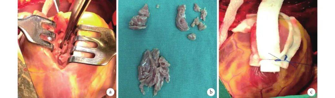
Surgical procedure: The patient was placed supine under general anesthesia; the chest was prepped and draped. Median sternotomy and pericardiotomy exposed the heart. After heparinization, aortic and bicaval cannulation with left ventricular venting established cardiopulmonary bypass. After cooling on bypass, the aorta was cross-clamped and HTK cardioplegia infused via the aortic root. With the heart arrested, ice slush was placed in the pericardial cavity for deep hypothermia. A pad under the heart base tilted the apex forward. Avoiding coronary territories, a 10 cm incision was made at the LV apex lateral and away from the LAD; retractors exposed the cavity, papillary muscles, and chordae (Figure 4a), matching the preoperative 3D model. Hypertrophied septum and LV anterior/lateral wall myocardium were resected layer by layer from apex toward base (Figure 4b), enlarging the apical cavity until the mitral leaflets and anterior/posterior papillary muscle groups were clearly visible. Cavity relief was satisfactory without mitral injury. The incision was closed with two felt strips along both sides and a long felt strip reinforcing the midline (Figure 4c). After confirming no bleeding or coronary injury, the aorta was unclamped, the heart restarted, temporary ventricular pacing wires placed, CPB weaned, pericardial and mediastinal drains left, and the chest closed layer by layer before ICU transfer. CPB time was 117 min; aortic cross-clamp time 57 min. The patient returned to the ward on postoperative day 2 and was discharged on day 7. Predischarge color Doppler echocardiography showed an enlarged apical cavity versus before (Figure 2); mid-to-lower septal thickness 12 mm; unobstructed mid-to-lower LV flow at 0.75 m/s; LV volume 45 mL; mild mitral regurgitation, area 1.0 cm2; no systolic anterior motion (SAM) of the anterior mitral leaflet. Symptoms improved with resolution of exertional dyspnea. No perioperative complications occurred; discharge was uneventful.
 Surgery
Figure 4 Intraoperative views
a: Apical incision exposing the cavity; asterisk marks myocardium to be resected; b: Resected myocardium; c: Felt-strip reinforcement of the apical incision
This study was approved by the Medical Ethics Committee of Guangdong Provincial People’s Hospital (Guangdong Academy of Medical Sciences), approval No. KY-Q-2022-035-01, with patient informed consent.
Discussion This is the first domestic report of apical-approach myectomy guided by 3D printing for hypertrophic cardiomyopathy. Hypertrophic cardiomyopathy is an autosomal dominant cardiomyopathy characterized anatomically by asymmetric left ventricular wall hypertrophy and is among the most common causes of sudden cardiac death in adolescents and athletes[1]. Pathologic hallmarks include asymmetric septal hypertrophy, left ventricular outflow tract obstruction, and SAM of the anterior mitral leaflet. Apical hypertrophic cardiomyopathy is uncommon, with higher prevalence in Asian than non-Asian populations (25% vs. 10%), typically presenting at ages 30–50 and more often in males[2-3]. Unlike typical HCM, apical HCM frequently features mid-ventricular obstruction and apical aneurysm. Early disease may be asymptomatic, but progressive diastolic dysfunction increases LV pressure load and left atrial size, leading in mid-to-late stages to chest tightness, dyspnea, and reduced exercise tolerance. Increased wall tension impairs coronary perfusion with microvascular disease, and imbalance between myocardial oxygen supply and demand can accelerate progression and aneurysm formation[4-5]. Beyond symptoms, imaging is characteristic. Transthoracic color Doppler echocardiography is the simplest direct modality; end-diastolic maximum wall thickness ≥15 mm distal to the papillary muscle insertion confirms diagnosis. Deep T-wave inversion (≥10 mm) is an ECG feature of apical HCM but does not distinguish coronary disease or other hypertrophic phenotypes. In a long-term follow-up of 105 apical HCM patients, only 47% had deep T-wave inversion[6]. Left ventriculography may show the “ace of spades” sign, and with aneurysm a “teardrop” appearance of high diagnostic value[7]. Aneurysm may also be confirmed by paradoxical motion on transthoracic echocardiography, CT showing continuous but thinned apical wall, or perfusion defects on single-photon emission computed tomography (SPECT). About 2% of HCM patients have aneurysms; compared with other phenotypes without aneurysm, prognosis is worse, with significantly higher risk of sudden death, thromboembolic events, sustained ventricular tachycardia, progressive heart failure, and other adverse cardiovascular events[8-10].
Extended myectomy via the aortic approach (Morrow procedure) is the traditional surgical treatment for HCM. For apical hypertrophy, resection is theoretically possible but deep, with narrow visualization and limited working space, making surgery very difficult and raising risks of septal perforation and injury to valves or coronaries. Some authors[11-12] have proposed apical-approach hypertrophic myectomy as a safe, effective option for apical hypertrophy, allowing ready exposure and management of hypertrophied myocardium, abnormal muscle bundles, and papillary muscles adherent to the wall, with good postoperative stroke volume and functional recovery. For complex cases with mid-ventricular obstruction, combined aortic and apical dual approaches also yield marked clinical benefit[13]. Another advantage of the apical approach is aneurysm management: the incision can be placed over the aneurysm for direct resection and thrombus clearance. Domestic reports of apical-approach HCM surgery remain limited; outcomes depend on operator experience, with common complications including incomplete relief of obstruction, septal perforation, and mitral injury. To address these issues, this case used 3D modeling and printing for accurate preoperative assessment of resection site and extent. Routine preoperative studies include transthoracic/transesophageal echocardiography, cardiac CT, and cardiac MRI, but 2D imaging lacks depth and still requires intraoperative exploration to reconstruct hypertrophied geometry before resection. Preoperative 3D modeling and printing allow intuitive, accurate localization of hypertrophied myocardium relative to papillary muscles, mitral valve, and aneurysm, and determination of incision site; planar cutting can further simulate intraoperative viewpoints and precisely plan resection extent and pathway within one field of view, enabling personalized preoperative planning and reducing difficulty. Finally, apical myectomy often relies on visual inspection or digital palpation to judge relief of obstruction, typically needing a larger incision with vague feedback; 3D modeling can annotate and quantify the shape and volume of myocardium to be resected for real-time comparison with the planned model to ensure effect.
This report provides practical support for apical-approach myectomy, reaffirming that apical resection is safe and effective for apical hypertrophic cardiomyopathy, and that 3D modeling and printing are important tools to guide complete relief of obstruction and prevent complications.
 Sailner Cardiovascular Medicine 3D Printing Joint Laboratory
Zhuhai Sailner Digital Medical and the Guangdong Provincial People’s Hospital Cardiovascular Institute formally signed an agreement to establish the “Cardiovascular Medicine 3D Printing Joint Laboratory,” a frontier medical research and application institute jointly built by hospital and industry. Centered on cardiovascular clinical and educational applications, the joint laboratory focuses on medical 3D applications and cutting-edge technology, selects distinctive topics, and pools both parties’ strengths for key technical research, striving for major application outcomes in medical 3D printing to drive related research, promotion, and adoption.
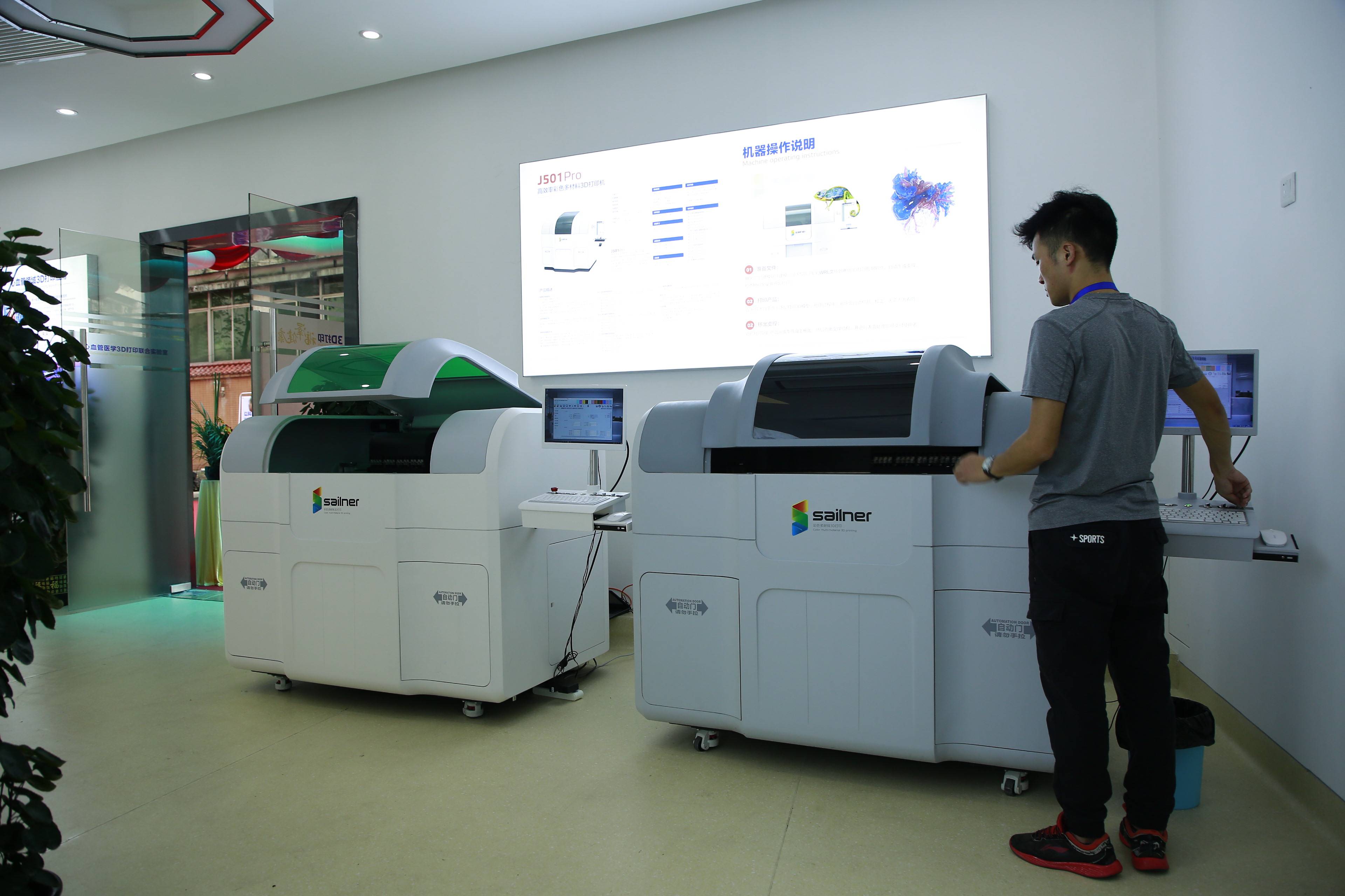 Sailner Digital Medical 3D printer
Most of this article is adapted from “Apical-approach myectomy guided by 3D printing for apical hypertrophic cardiomyopathy: a case report” in the Chinese Journal of Clinical Thoracic and Cardiovascular Surgery,
Tan Tong, Wei Peijian, Liu Jian, Zang Xin, Zhu Wei, Wu Hongxiang, Liu Yanjun, Li Xiaoyi, Zhuang Jian, Guo Huiming